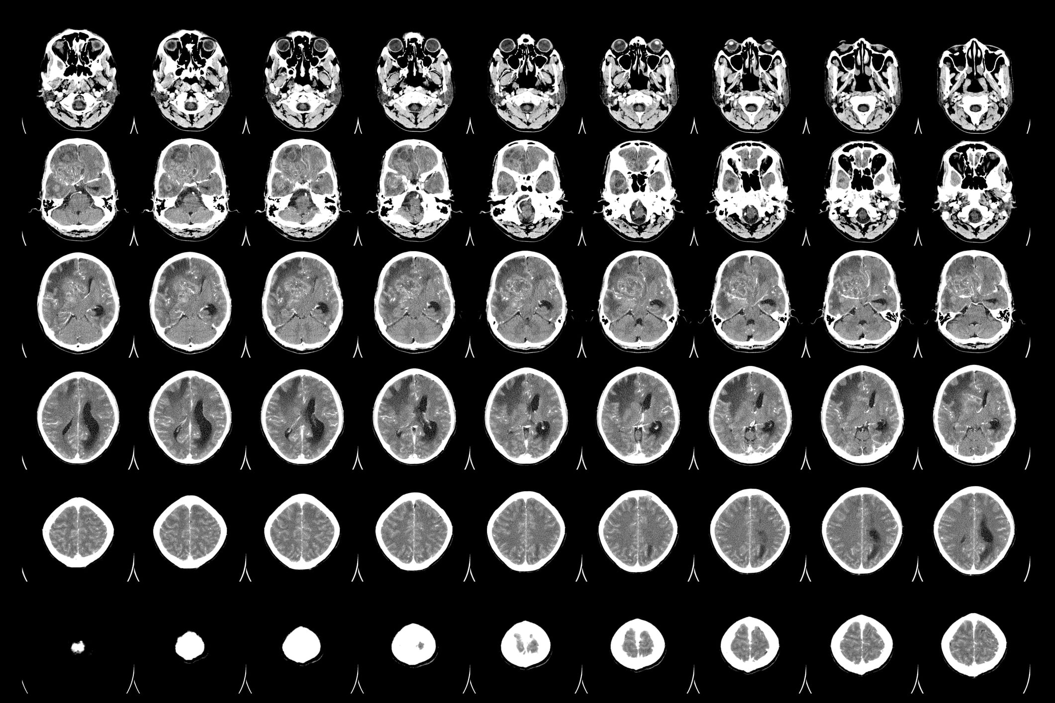
In an unprecedented move to transform neurosurgery and improve the safety and efficacy of brain tumour removal, leading institutions at Imperial College London are jointly developing innovative ‘bionic eye’ technology. This groundbreaking collaboration involves the Brain Tumour Research Centre of Excellence at Imperial College and the Hamlyn Centre, one of the Institute of Global Health Innovation’s key research facilities.
The revolutionary ‘bionic eye’ will leverage highly sophisticated imaging technology to clearly distinguish between healthy and diseased brain tissue. This cutting-edge approach is geared towards the removal of as much tumour tissue as possible while preserving critical brain matter.
Surgeons currently grapple with formidable challenges in extracting certain tumours, such as glioblastoma (GBM), which are diffuse in nature. These cancerous forms infiltrate healthy brain tissue without discernible boundaries, making surgical intervention both complex and perilous.
The technology under development will utilize a vast database of images. Work is already afoot to train a precise algorithm that can differentiate between tumour and healthy brain tissue. Furthermore, this algorithm will identify critical areas of the brain responsible for essential functions like speech. These advancements will culminate in the development of a ‘bionic eye’ apparatus that can be integrated into a surgical microscope, providing neurosurgeons with vital real-time insights during operations.
Dr. Karen Noble, Director of Research, Policy, and Innovation at the Brain Tumour Research Centre of Excellence, has lauded the project, stating, “This exciting project is at the cutting edge of surgical research, and we congratulate the team at Imperial on their progress so far.”
The promising technology brings more than precise distinction between diseased and healthy tissue; it also provides invaluable information for avoiding areas of the brain vital for functions like speech and mobility. As a result, surgeons can be more proactive in eradicating tissue surrounding the tumour without inflicting permanent damage.
“This is a giant leap towards safer, more effective brain surgery,” says Dr. Noble. “Not only does it pave the way for a more targeted removal of the tumour, but it also preserves essential brain functions, significantly enhancing patients’ quality of life post-surgery.”
The ongoing work represents a significant stride in medical innovation, marking a hopeful future in the world of neurosurgery and brain tumour research. Collaborative efforts between Imperial College’s top research centres reveal the potent possibilities of inter-disciplinary work in the realm of medical science.
Dr Giulio Anichini, a clinical research fellow funded by Brain Tumour Research, said: “This is an exciting development for glioma and GBM patients, and potentially to be used in other areas of neurosurgery. It allows us to be much more precise in removing diseased tissue versus healthy tissue. It also means we can determine areas of the brain which are crucial to vital functions such as speech and mobility, and those where we’d be more proactive in removing tissue around the tumour without causing lasting damage.”
This technological advancement, upon completion and subsequent integration into regular surgical procedures, has the potential to redefine the frontiers of neurosurgical treatment, saving countless lives and improving post-surgical recovery and functionality.
The medical community and those affected by brain tumours eagerly await further updates on this transformative project. The fusion of artificial intelligence with advanced imaging presents an optimistic horizon in the complex world of neurosurgery, setting new standards for precision, safety, and overall success.
Subscribe
Sign-up to receive our newsletter


